
The Cholesterol Conundrum: Ancestral Fats, Modern Arteries, and the Biology of Vascular Repair
For nearly two million years, ancestral human physiology thrived on the nutrient-dense fats of wild game, yellow bone marrow, and coastal marine life. Yet within a single generation in the mid-twentieth century, the foundational building blocks of human cell membranes were rebranded as dietary poison. Today, the debate over saturated fat, low-density lipoproteins (LDL), and cardiovascular disease is reaching a breaking point.
To understand whether animal fat causes arterial plaque—or whether our modern industrial environment is destroying the vascular highway—we must examine evolutionary anthropology, the collapse of pre-industrial dietary norms, the flawed roots of modern nutritional dogma, and the delicate biomechanics of the vascular wall.
1. What We Actually Ate: Two Million Years of Pleistocene Fuel
The human gastrointestinal tract and metabolic machinery evolved during the Middle and Late Pleistocene—an epoch defined by glacial cycles and vast herds of terrestrial megafauna. Hominins such as Homo erectus, Neanderthals, and early Homo sapiens did not eat the marbled muscle steaks found in modern supermarkets. Wild game muscle is exceptionally lean, often carrying less than 3% intramuscular fat.
To survive without succumbing to protein toxicity (historically known as “rabbit starvation”), hominins developed specialized behavioral strategies to harvest concentrated anatomical fat caches:
- Yellow Bone Marrow: Long before hominins were apex hunters, early humans used hammerstones to access yellow marrow cavities. Marrow is roughly 75% to 85% fat, dominated by monounsaturated oleic acid (18:1)—the same fat celebrated in olive oil.
- Brain & Central Nervous Tissue: The cranial vault was fractured for structural lipids rich in preformed docosahexaenoic acid (DHA, 22:6 n-3) and arachidonic acid (ARA, 20:4 n-6).
- Visceral & Subcutaneous Deposits: Mesenteric fat, kidney suet, and thick subcutaneous fat pads from giant herbivores provided stable saturated fatty acids (palmitic and stearic acid).
Primary scavenged lipid cache: 75%–85% pure fat.
Dominant Lipid: Oleic Acid (MUFA ~60%).
Fluid / Bioavailable
Structural tissue: 60% dry weight structural lipids.
Dominant Lipid: Preformed DHA & ARA.
Encephalization Fuel
Megafaunal kidney fat and subcutaneous pads.
Dominant Lipid: Stearic & Palmitic Acid.
High Oxidative Stability
Evolutionary Omega Ratio: Maintained strictly between 1:1 and 2.5:1. Zero industrial seed oils.
Figure 1: The Evolutionary Lipid Template. For over 1.5 million years, human dietary fat came from concentrated anatomical organs rather than marbled feedlot muscle.
2. The Historical “Experiment”: 19th-Century Subsistence Without Heart Attacks
Skeptics often claim ancestral examples are irrelevant because early humans supposedly died too young to develop cardiovascular disease. However, historical demographic records from the 18th and 19th centuries provide a clear rebuttal.
Consider the rural Irish cottier prior to the Great Famine of 1845. An adult agricultural laborer consumed between 8 and 14 pounds (3.5 to 6 kg) of potatoes daily, paired with:
- Farmhouse Churn Buttermilk: Drinking 1 to 3 pints daily of unpasteurized cultured buttermilk, retaining milk fat globule membranes (MFGM), butyric acid, and fat-soluble vitamins.
- Salted Pig Scrap & Lard: Scrap fatback and bacon dripping (rich in monounsaturated oleic and saturated stearic acids) were boiled alongside the tubers.
- Dried Salt Herring: Consumed widely across coastal and inland parishes, providing intact marine omega-3s.
• 8–14 lbs boiled potatoes / day
• ~3,500–4,200 calories
• Zero industrial sugar or refined flour
Burned via Heavy Labor
• 1–3 pints churn buttermilk + pig lard
• ~25–45g animal lipid / day (~8% of calories)
• Vehicle for fat-soluble vitamins (A, D, K2)
Zero Industrial Processing
Clinical Outcome: Occlusive coronary heart disease was virtually unrecorded in 19th-century autopsy records. Acute myocardial infarction only surged after 1920.
Figure 2: The Pre-Industrial Baseline. Traditional populations lived on unrefined starches paired with natural animal fats for generations without epidemics of occlusive coronary thrombosis.
3. The 1950s Pivot: Panic, Ancel Keys, and Corporate Capture
Between 1920 and 1950, occlusive coronary heart attacks exploded across the Western world. When US President Dwight D. Eisenhower suffered a sudden heart attack in 1955, clinical medicine faced a public crisis. An answer was demanded immediately.
Physiologist Ancel Keys stepped forward with the Diet-Heart Hypothesis: saturated fat raises serum cholesterol, cholesterol builds plaque, and plaque clogs coronary arteries. Keys popularized this concept through the Seven Countries Study, presenting a neat line between dietary saturated fat and coronary death.
Figure 3: The Mid-Century Consensus. The intuitive “plumbing analogy” assumed linear causality, mistaking a surrogate blood marker for the total cause of vascular pathology.
In 1948, Procter & Gamble contributed over $1.5 million in modern purchasing power to the American Heart Association (AHA). By 1961, the AHA officially recommended replacing traditional butter, lard, and tallow with polyunsaturated vegetable oils and margarine.
• Beef Tallow, Butter, Lard
• High heat stability, resistant to rancidity
• Rich in natural stearic and oleic acid
Eaten for Millennia
• Soybean, Corn, Cottonseed Oils, Crisco
• Extracted with chemical solvents and bleached
• Highly prone to lipid peroxidation; loaded with trans fats (1950–2000s)
Promoted as “Heart-Healthy”
Figure 4: The Industrial Food Shift. Low-margin natural animal fats were displaced by highly profitable, chemically deodorized seed oils and trans-fat-laden stick margarines.
4. The Road and the Subsidence: The True Anatomy of Arterial Plaque
The persistent error of standard lipidology is treating circulating cholesterol as an independent poison. Lipids do not float naked in the bloodstream; they are hydrophobic molecules packaged inside hydrophilic transport vehicles known as lipoproteins (chylomicrons, VLDL, IDL, LDL, and HDL).
Blaming low-density lipoprotein (LDL) for heart disease because it is found inside plaques is equivalent to blaming emergency vehicles for causing a pile-up on a washed-out highway. To understand why plaques form, we must look at the road itself: the vascular endothelium.
ApoB-containing lipoproteins (LDL, VLDL) circulate freely, delivering vital sterols to peripheral tissues without adhering.
A dense, negatively charged gel matrix of proteoglycans and glycoproteins that physically bars lipoproteins from touching endothelial junctions.
Endothelial tight junctions remain closed; subendothelial intimal tissue remains protected and sterile.
Figure 5: The Intact Vascular Highway. An intact endothelial glycocalyx repels passing lipoproteins regardless of ambient particle count.
Step 1: The Non-Stick Glycocalyx
A healthy blood vessel is lined by the endothelial glycocalyx—a brush-like, gel matrix of glycoproteins and proteoglycans. When this coating is intact, passing ApoB-containing lipoproteins bounce harmlessly off the arterial wall, cycling between peripheral tissues and hepatic clearance receptors.
Step 2: Road Subsidence (Endothelial Breakdown)
When an individual consumes a modern diet high in refined carbohydrates, high-fructose syrups, and unstable omega-6 seed oils, three destructive processes occur:
- Glycocalyx Stripping: Sharp spikes in postprandial blood glucose activate shedding enzymes that dissolve the protective non-stick matrix.
- Oxidative Derangement: Circulating linoleic acid integrates into lipoprotein membranes, leaving them vulnerable to lipid peroxidation and producing reactive aldehydes (such as 4-HNE and MDA).
- Endothelial Retraction: Loss of bioavailable nitric oxide (eNOS decoupling) causes endothelial cells to retract, exposing microscopic gaps in the basement membrane.
Figure 6: Arterial Subsidence. Damage to the vascular roadbed permits particle entrapment and initiates plaque formation.
Step 3: Trapping and the Immune Response
Once oxidized or glycated, ApoB particles can no longer bind properly to liver LDL receptors for clean recycling. They slip through endothelial fissures, bind to subendothelial proteoglycans, and become trapped. The immune system recognizes these oxidized lipids as foreign threats and dispatches monocytes.
Smooth muscle cells migrate to lay down collagen, sealing the toxic lipid core away from the bloodstream.
Monocytes transform into macrophages, engulfing trapped, oxidized lipids until immobilized.
Accumulation of ruptured foam cells, cholesterol crystals, and inflammatory debris. Rupture of this cap causes acute heart attacks.
Figure 7: Plaque Genesis. Plaque is an active, living wound-healing response to persistent vascular insult.
5. Arteries vs. Veins: The Hemodynamic Smoking Gun
If circulating cholesterol concentration alone were the primary cause of atherosclerosis, plaque would develop uniformly throughout the entire circulatory system. Yet it does not.
• Pressure: Low (~5–15 mmHg)
• Flow Dynamics: Smooth, non-pulsatile laminar flow
• Lipid Concentration: Identical circulating ApoB/LDL
Atherosclerosis Rate: ~0%
• Pressure: High (~120/80 mmHg)
• Flow Dynamics: Severe shear stress at bifurcations
• Lipid Concentration: Identical circulating ApoB/LDL
Plaque Concentrates Here
The Surgical Proof: Veins only develop atherosclerosis when harvested and grafted into the high-pressure arterial circulation during coronary bypass surgery.
Figure 8: The Hemodynamic Reality. Mechanical shear stress and high pressure are required prerequisites for plaque formation.
Blood circulating through veins contains the same concentration of LDL, ApoB, and Lp(a) as blood in arteries. However, veins virtually never develop atherosclerosis. Plaque occurs almost exclusively in the arterial tree—specifically at sharp bifurcations where blood flow becomes turbulent, exposing the endothelial lining to chronic mechanical shear stress.
6. Navigating the Transition: Why Going Carnivore in Your 50s or 60s Requires Caution
Given that animal fats are an evolutionary baseline and refined carbohydrates and seed oils drive vascular inflammation, many adults choose to adopt a strict low-carbohydrate or carnivore diet. However, transitioning abruptly in mid-to-late life introduces specific physiological variables that require careful management.
Zero carb intake forces the liver into systemic lipid trafficking, dispatching high numbers of VLDL/LDL to shuttle fat fuel.
Rapid sodium/water flushing spikes cortisol and catecholamines, temporarily raising pulse pressure on delicate plaques.
Sudden drop in systemic inflammation triggers enzymatic remodeling of thin, vulnerable fibrous caps before they can calcify.
Figure 9: The Acute Transition Surge. Sudden dietary shifts can introduce mechanical and biochemical friction across preexisting vascular plaques.
After five or six decades on a standard Western diet, the arterial wall is rarely in a pristine state. Many individuals carry subclinical, asymptomatic plaques. Some of these are stabilized by thick fibrous caps, while others are “vulnerable plaques”—soft lipid pools covered by a thin, fragile layer of collagen.
The Three Acute Transition Stressors:
- The ApoB Particle Surge: When carbohydrate intake drops to zero, the liver shifts entirely to systemic lipid trafficking, dispatching large numbers of VLDL and LDL particles to deliver energy to peripheral tissues. In lean or metabolically active individuals, ApoB particle counts can double or triple within weeks.
- Electrolyte Flushing and Sympathetic Spikes: As insulin falls, the kidneys excrete sodium and water rapidly. This sudden fluid shift can trigger a compensatory release of cortisol, epinephrine, and norepinephrine, temporarily raising blood pressure and shear stress across existing plaques.
- Plaque Remodeling Dynamics: As systemic inflammation drops, matrix metalloproteinases begin remodeling the extracellular matrix of arterial walls. If an inflamed, thin-capped plaque undergoes rapid shifts in lipid flux and mechanical stress before it can fibrose and calcify securely, the risk of an acute rupture increases.
7. The Pharmacological Bridge: Balancing Biology with Prudence
For individuals over 50 with documented vascular disease or elevated Coronary Artery Calcium (CAC) scores (> 100–400), an abrupt surge in circulating particles across fragile arterial walls carries practical risks. In this specific window, using low-dose statin therapy as a temporary pharmacological bridge provides a rational, physiological safety measure[span_0](start_span)[span_0](end_span)[span_1](start_span)[span_1](end_span).
• Thin, fragile collagen caps on existing plaques[span_2](start_span)[span_2](end_span)[span_3](start_span)[span_3](end_span)
• Endothelial junctions inflamed / slowly healing[span_4](start_span)[span_4](end_span)[span_5](start_span)[span_5](end_span)
• High risk of rupture under sudden particle or pressure surges[span_6](start_span)[span_6](end_span)[span_7](start_span)[span_7](end_span)
Fragile Arterial State
• Pleiotropic effect: Promotes thickening of protective fibrous caps[span_8](start_span)[span_8](end_span)[span_9](start_span)[span_9](end_span)
• Upregulates hepatic LDL clearance receptors[span_10](start_span)[span_10](end_span)[span_11](start_span)[span_11](end_span)
• Blunts particle surges while glycocalyx repairs under zero glycation[span_12](start_span)[span_12](end_span)[span_13](start_span)[span_13](end_span)
Protective Bridge
Long-Term Strategy: Once systemic inflammation normalizes (hs-CRP < 0.5 mg/L, fasting insulin < 5 uIU/mL), ongoing pharmacological need can be safely re-evaluated with your clinician[span_14](start_span)[span_14](end_span)[span_15](start_span)[span_15](end_span).
Figure 10: The Pharmacological Bridge Mechanism. A short-term statin bridge stabilizes fragile plaques and manages particle volume while the vascular endothelium heals on an ancestral diet[span_16](start_span)[span_16](end_span)[span_17](start_span)[span_17](end_span).
While statins have trade-offs—such as inhibiting the HMG-CoA reductase pathway and dampening endogenous Coenzyme Q10 (CoQ10) production—their immediate clinical value lies in their pleiotropic, anti-inflammatory effects[span_18](start_span)[span_18](end_span):
- They inhibit vascular cell adhesion molecules and calm macrophage activation within existing plaques[span_19](start_span)[span_19](end_span).
- They promote the rapid thickening and cross-linking of the protective collagen fibrous cap, reducing the risk of plaque rupture[span_20](start_span)[span_20](end_span)[span_21](start_span)[span_21](end_span).
- They upregulate hepatic LDL clearance receptors, preventing extreme particle surges while the vascular glycocalyx repairs itself in an environment free from dietary glycation[span_22](start_span)[span_22](end_span).
Once systemic inflammation drops (demonstrated by an hs-CRP < 0.5 mg/L, a normalized fasting insulin level, and an optimal triglyceride-to-HDL ratio) and the vascular endothelium stabilizes, the patient and physician can re-evaluate long-term pharmacological support from a position of metabolic strength[span_23](start_span)[span_23](end_span)[span_24](start_span)[span_24](end_span).
Summary Protocol: Phasing the Transition Safely
| Phase | Timeframe | Clinical Action & Nutrition Target |
|---|---|---|
| 1. Diagnostic Verification | Baseline (Week 0) | Obtain a low-dose Coronary Artery Calcium (CAC) scan and advanced lipid panel (ApoB, hs-CRP, Fasting Insulin, HbA1c). Establish true baseline vascular burden. |
| 2. Industrial Elimination | Weeks 1–3 | Eliminate 100% of industrial seed oils (soybean, canola, corn, sunflower) and refined sugars. Retain whole-food carbohydrates (sweet potatoes, fruit) to prevent acute electrolyte flushing. |
| 3. Carbohydrate Taper | Weeks 4–6 | Step down carbohydrates systematically (150g → 100g → 50g daily). Supplement baseline electrolytes (sodium chloride, potassium citrate, magnesium malate) to support stable vascular tone. |
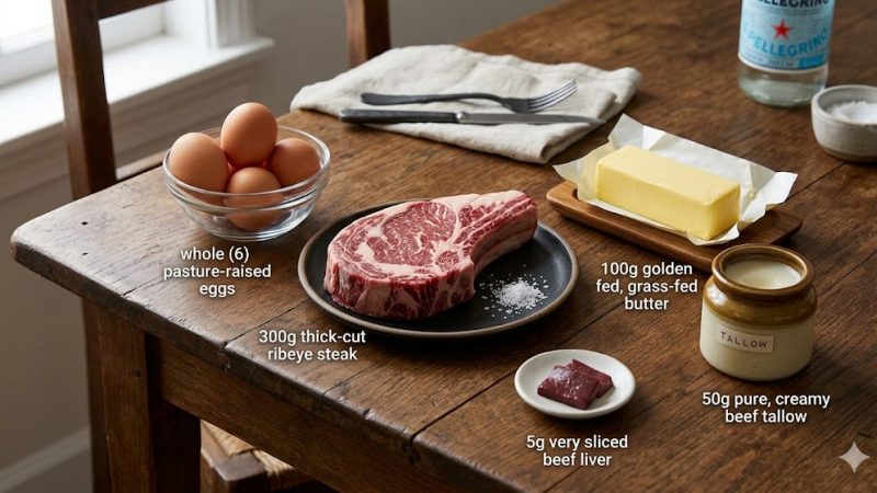
| 4. Whole Animal Transition | Weeks 7+ | Transition fully into nutrient-dense ruminant meats, tallow, pastured eggs, liver, and marrow. Re-test hs-CRP, ApoB, and metabolic markers at 90 days. |
Cardiovascular disease is not the product of an ancestral fat that sustained human evolution for two million years. It is the consequence of an engineered, modern food supply that degrades the arterial lining, alters lipid chemistry, and leaves our vascular repair systems overwhelmed. By understanding the mechanical reality of the vascular wall, we can move past outdated dietary dogma and rebuild long-term cardiovascular resilience safely.